Congenital heart defects
Ventricular septal defect (VSD)
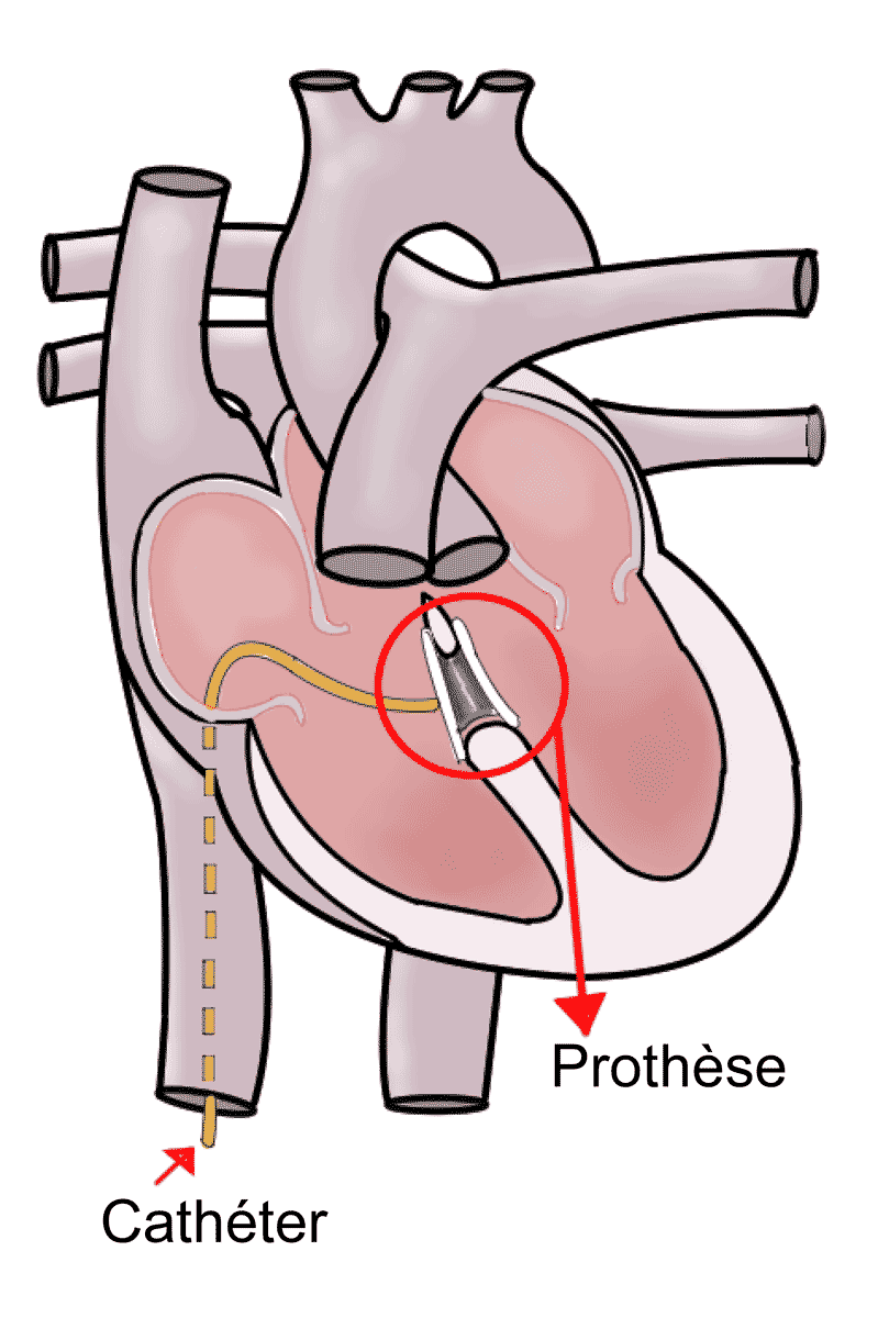
Closure of a ventricular septal defect (VSD), an abnormal opening between the two ventricles.
What is it?
A ventricular septal defect (VSD) is an opening in the wall that separates the two ventricles, the lower chambers of the heart. Blood passes from the left ventricle to the right ventricle and reaches the lungs in excess. It is the most common congenital heart defect. Small defects often close on their own; larger ones are closed surgically, generally within the first months of life.
Step by step
- 1
An echocardiogram shows the size and position of the defect and its effect on the lungs. It determines when the operation should take place.
- 2
The operation is performed under general anaesthesia: your child remains asleep for the whole procedure.
- 3
The surgeon reaches the heart through an opening of the sternum, the bone in the middle of the chest.
- 4
Cardiopulmonary bypass, a machine that temporarily takes over the work of the heart and lungs, allows the repair to be done on a still heart.
- 5
The defect is closed with a patch, usually working through the right atrium and the tricuspid valve, without cutting into the ventricular muscle.
- 6
An echocardiogram performed in theatre confirms the closure. The heart resumes its activity and your child is transferred to paediatric intensive care for continuous monitoring.
Benefits and expected outcome
The aim is to stop the passage of blood between the two ventricles and bring blood flow to the lungs back to normal. This protects the lung arteries from excessive pressure and reduces the workload on the heart. Most children who are operated on then regain weight gain and activity appropriate for their age.
Recovery and follow-up
Hospital stay usually lasts a few days, the first of them in paediatric intensive care. Feeding is resumed as soon as your child's condition allows, and the heart medicines started before the operation are often reduced and then stopped. At home, the scar is checked and activity is resumed gradually. Echocardiographic follow-up is scheduled to verify the closure and the heart rhythm.
This page is for information and does not replace a medical consultation. Each situation is different and is assessed individually.
Appointment
An opinion on your situation?
Don't wait to talk about it: book an appointment.