
Cardiac tumour surgery
Cardiac tumours
Surgical removal of a cardiac tumour, most often benign.
What is it?
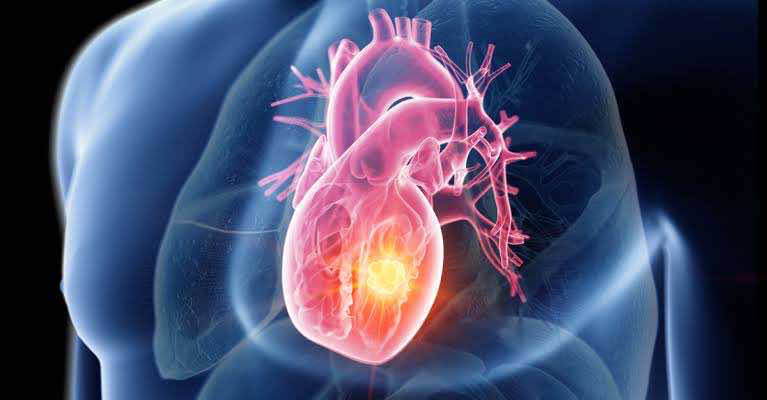
A cardiac tumour is a mass that develops inside a chamber of the heart or on its wall. In the large majority of cases it is benign; the most common type, a myxoma, usually sits in the left atrium. Depending on its size and position, it can obstruct blood flow or interfere with a valve. The standard treatment is surgical removal of the mass.
Step by step
- 1
The diagnosis is confirmed by an echocardiogram (an ultrasound image of the heart), completed if needed by a CT scan or an MRI. These examinations show the size of the mass and where it is attached.
- 2
The operation is performed under general anaesthesia: you remain asleep throughout.
- 3
The surgeon reaches the heart through a vertical opening of the sternum, the bone in the middle of the chest.
- 4
Cardiopulmonary bypass, a machine that temporarily takes over the work of the heart and lungs, allows the cardiac chamber to be opened and the surgery to be carried out on a still heart.
- 5
The mass is removed in full, together with its base of attachment. If required, the wall is repaired with a patch of pericardium, the membrane surrounding the heart.
- 6
The heart resumes its activity, the sternum is closed and you are transferred to intensive care for continuous monitoring. The removed mass is analysed in the laboratory.
Benefits and expected outcome
The aim of the operation is to remove the mass and restore normal blood flow through the heart. Most patients see an improvement in the symptoms that led to the diagnosis, such as breathlessness or palpitations. Microscopic analysis of the removed specimen establishes the exact nature of the tumour and guides follow-up.
Recovery and follow-up
Hospital stay usually lasts a few days, the first of them in an intensive care unit. Getting up and walking are resumed early, with the help of physiotherapists. The sternum takes about two months to heal: driving, lifting and effort with the arms are restricted during this period. A follow-up echocardiogram is scheduled to check that the tumour has not returned.
This page is for information and does not replace a medical consultation. Each situation is different and is assessed individually.
Appointment
An opinion on your situation?
Don't wait to talk about it: book an appointment.