Congenital heart defects
Atrial septal defect (ASD)
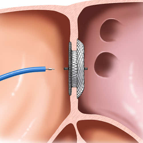
Closure of an atrial septal defect (ASD), an abnormal opening between the two atria.
What is it?
An atrial septal defect (ASD) is an opening in the wall that separates the two atria, the upper chambers of the heart. Some blood passes from the left atrium to the right atrium, which increases the flow to the right side of the heart and to the lungs. It is present from birth and is often found when a murmur is heard or during an echocardiogram. Small defects may close on their own in the first years; the others are closed either through a catheter or by surgery.
Step by step
- 1
An echocardiogram shows the size, position and rims of the defect. It makes it possible to choose between catheter closure and surgical closure.
- 2
The operation is performed under general anaesthesia: your child remains asleep for the whole procedure.
- 3
The surgeon reaches the heart through an opening of the sternum; in some cases a smaller incision on the side of the chest can be used.
- 4
Cardiopulmonary bypass, a machine that temporarily takes over the work of the heart and lungs, allows the atrium to be opened and the repair to be done on a still heart.
- 5
The defect is closed with direct stitches or with a patch of pericardium, the membrane surrounding the heart.
- 6
An echocardiogram performed in theatre confirms the closure. The heart resumes its activity and your child is transferred to paediatric intensive care for continuous monitoring.
Benefits and expected outcome
The aim is to stop the abnormal passage of blood between the two atria. The right side of the heart and the lungs then no longer receive excess flow, which reduces the long-term risk of enlargement of the right chambers, rhythm disturbances and pulmonary hypertension. Most children then return to normal activity and growth.
Recovery and follow-up
Hospital stay usually lasts a few days, the first of them in a monitoring unit. Pain is treated, and feeding and walking are resumed as soon as your child's condition allows. School and sport are taken up again gradually, following the instructions given at discharge. A follow-up echocardiogram is scheduled to check the closure and the working of the heart.
This page is for information and does not replace a medical consultation. Each situation is different and is assessed individually.
Appointment
An opinion on your situation?
Don't wait to talk about it: book an appointment.